

ORTHOMANUAL VETERINARY TREATMENT OF A DACHSHUND WITH POSTERIOR PARALYSIS
By J. Heukels and D. C. Aharon Presented at the ESVN symposium in Paris, 2013.
Summary
An 8 year old Dachshund presented with symptoms of acute posterior paralysis and loss of bladder control. The patient was clinically diagnosed as having thoracolumbar intervertebral disc disease (TLDD), grade IV. At the request of the owner, no additional diagnostic testing was performed, with a non-surgical treatment route elected. The dog was given orthomanipulation, followed by two weeks of cage rest. Two weeks later, the dog could walk independently and had regained control of his bladder, but was still paraparetic. Six weeks after the treatment, the dog had clinically and neurologically recovered. Orthomanipulation was found in this case to be an effective, non-invasive and economical treatment option for dogs with TLDD.
Introduction
Acute back problems related to pathology of the intervertebral disc are commonly seen in the chondrodystrophic dog breeds.7,20,24 Hansen type 1 intervertebral disc disease (IVDD) is characterized by chondroid degeneration prior to extrusion or protrusion of the nucleus pulposus or annulus fibrosus.5,8,20,35 Extrusion or protrusion can cause compression and damage to the spinal cord and nerve roots.20,32 The thoracolumbar region of the spine is predisposed to IVDD,24,35 and the condition is known as thoracolumbar intervertebral disc disease (TLDD).
In the initial phase the extruded intervertebral disc causes damage to the spinal cord due to compression and contusion. Disruption of the cell membranes and blockage of spinal blood supply and drainage lead to ischemic necrosis of neurons and glia cells and damage to myelin and axons. This primary phase sets a series of secondary mechanisms in motion, causing the affected tissue area to expand. Cell death is the result of rapid changes in intracellular ion concentrations, excitotoxicity (excessive neurotransmitter stimulation), destruction of the microvascular bed, production of free radicals and inflammation.20,32
The clinical manifestation of TLDD is dependent on the severity of damage to the white and grey matter of the spinal cord, caused by primary and secondary trauma. The symptoms vary greatly between individuals, and may include local pain, increased paraspinal muscle hypertonicity and pelvic limb neurological deficits.2,7,20 Pain is a result of irritation of and pressure on the meninges and nerve roots.10,20,24,35
Classification of the neurological state is dependent on the severity of the deficit (see table).20,24,34 There are different treatment options for clinical TLDD: conservative, i.e., cage rest and anti-inflammatory medication; non-surgical, i.e., physical therapy23,26 and acupuncture21; and surgical.7,20,24,26,28,32/ Manual treatment methods, such as orthomanipulation, are regularly used to treat back problems in humans 4,39 while orthomanual techniques have also been applied in veterinary medicine in recent years.1
Orthomanual medicine is based on the principle that intervertebral disc degeneration causes vertebral instability. That instability can induce misalignment of consecutive vertebrae. Vertebral misalignment, instability and intervertebral disc degeneration cause extrusion and/or protrusion of the nucleus pulposus or the annulus fibrosus. Spinal cord compression and trauma due to kinetic energy from the extruded intervertebral disc can cause pain and neurological deficits.39
It is theorized that correcting the misalignment of the vertebrae diminishes pressure on the intervertebral disc. This creates an environment that facilitates recovery and improvement of the neurological state.1
| Grade | Neurological state | Proprioception | Bladder control | Deep pain perception |
|---|---|---|---|---|
| I | Pain | + | + | + |
| II | Paraparesis | - | + | + |
| III | Paralysis | - | + | + |
| IV | Paralysis | - | - | + |
| V | Paralysis | - | - | - |
Case description
Characteristics and symptoms An 8-year-old male Dachshund, weighing 12.6 kilograms, was presented at the Practice for Veterinary Orthomanual Medicine in Noorden (the Netherlands) with the following complaints: pelvic limb paralysis and back pain. Symptoms had appeared a day earlier and were of acute onset. On the first day of treatment the dog was given 50 mg prednisolone sodium succinate (Solu-delta- cortef), subcutaneously administered. Urinary catheterization was performed to relieve urinary retention. The owner was instructed to perform urinary catheterization four times a day at home.
Two years earlier, the dog had fully recovered from an acute spinal problem with grade IV pelvic limb paralysis after orthomanual treatment. In addition, the dog had been successfully treated with non-steroidal anti- inflammatory drugs (Rimadyl, 1 dd 50 mg for 10 days) and an on-leash revalidation regime for lameness of the right hind limb, clinically diagnosed as hip pain.
Clinical findings The dog was presented with posterior paralysis. During the general clinical examination no irregularities were found. An orthopaedic examination was carried out. The spinal reflexes were tested with the patient recumbent on both the right and left sides. The right hip was painful and could be dislocated with abduction. Muscle volume and tonicity were normal in all limbs. The M. Extensor carpi radialis reflex (C7-Th2), triceps reflex (C6-Th1) and withdrawal reflex (C6-Th2) of both front limbs were intact. The patellar reflex (L3/4-L6), tibialis reflex (L6-S1) and withdrawal reflex (L5-S1) of both hind limbs were intact.20,38 The perineal reflex (S1-S3) was intact. Superficial and deep pain perception were intact in all limbs. The knuckling reflex (proprioceptive positioning) was absent in both pelvic limbs.33 The bladder did not evacuate naturally, or with manual pressure on the abdomen.
Orthomanual examination of the back.
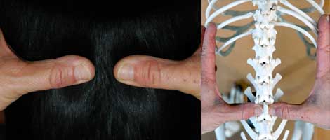
Neuroanatomic localization and differential diagnosis Summarized findings of the neurological examination were the following: symmetrically absent proprioception of the pelvic limbs, and intact spinal reflexes of all limbs. Impaired proprioception is an upper motor neuron (UMN) symptom and indicates a spinal cord lesion cranial to the segment of innervation of the relevant limb. Urinary retention with a bladder that has difficultly in evacuating, or cannot be evacuated using pressure, indicate a UMN micturition disorder. The normal spinal reflexes mean that these reflex arcs and the corresponding spinal cord segments are intact. Therefore, the lesion must be located between Th3 and L3, and classifies as grade IV. Differential diagnoses of acute Th3-L3 spinal cord lesions are: TLDD, trauma and fibrocartilaginous infarction or haemorrhage.9 Complementary diagnostics with myelography, computed tomography (CT) or magnetic resonance imaging (MRI)6,7,24 and subsequent surgery were discussed with the owner and advised. The owner elected against performance of further diagnostics and chose instead a non-surgical treatment route. Orthomanual examination and treatment Orthomanual examination of the back and neck was performed with the dog standing squarely in front of the veterinarian. An assistant supported the patient by placing both hands under the trunk to keep it steady. The veterinarian palpated the entire vertebral column from tail to head, by placing the thumbs on either side of the vertebral dorsal spinous process. Vertebral misalignments were detected by visual inspection of the position of the thumbs and by palpation. Asymmetry, local pain, paraspinal muscle tone and muscular atrophy were noted. The examination revealed local pain upon palpation of the back at the level of the thoracolumbar junction. Furthermore, misalignments of Th11, Th12 and Th13 were identified and corrected. A misaligned vertebral position was corrected by slightly elevating the vertebral body with the thumb and then applying a gentle thrust to the spinous process, thereby pushing it into the correct position.1 Typically, very little force is required to correct a misaligned vertebra, because the pressure is applied in the direction of the anatomically optimal position and function.39 A strict regime of cage rest was prescribed for a period of 14 days.
Progression and prognosis
At follow-up appointments, the orthopaedic, neurological and orthomanual examinations were repeated. At the two-week follow-up the dog was ambulatory with posterior paresis. The knuckling reflex was normal in the right hind leg, and it was delayed in the left hind leg. Furthermore, the dog was pain-free and had regained full bladder control. Over the subsequent four-week revalidation period, rest and a managed mobility regime was prescribed, i.e., gradually introducing more freedom of movement, alongside intervals of cage rest.24,35 At the six-week follow-up, the dog’s gait was normal and proprioception was intact in both pelvic limbs (neurological grade 0). The orthomanual treatment was therefore considered successfully completed and concluded.
Discussion
The probable diagnosis of TLDD is often based on patient characteristics, history, symptoms and the clinical examination. Considering the neuroanatomical localization and acute onset, TLDD was the most likely diagnosis for this patient.32 The typical age at which the first symptoms of TLDD appear is 3-7 years.7,20 This corresponds with the age at which this dog initially experienced back problems. A year later, the dog was seen again for lameness of the right hind leg. At that time mild recurrence of TLDD could not be entirely ruled out. However, there were no clinical indications of this. The dog did not have back pain, there was no neurological deficit and no vertebral misalignments. Recurrence of TLDD is a well-known phenomenon, regardless of treatment method.7,12,28 After surgical treatment, 2%-15% of patients experience recurrence. In Dachshunds, the chance of recurrence is 3-10 times higher.12,15,19,31 This usually concerns extrusion or protrusion of another intervertebral disc. After conservative treatment, 30%-50% of patients experience recurrence.20,24 No data is available on recurrence incidence after orthomanual treatment. Experience from practice suggests that the average time between a first episode and recurrence is longer following orthomanual treatment than after surgery. A possible explanation for this is that surgery eliminates the compression only locally. With orthomanual treatment, all misalignments in the spine are treated, thus including those in which subclinical IVDD may be present. A fibrocartilaginous infarction (FCI) is the primary differential diagnosis for this patient, though this is not common in the breed, and an FCI typically causes asymmetrical deficits.11,17 Complementary diagnostics are needed for confirmation of the diagnosis, but these bring added costs for the owner and risks for the patient (i.e., if anaesthesia is required).7,10,24 Intravenous administration of a high dose (30 mg/kg) of prednisolone sodium succinate within 8 hours of onset of signs of TLDD could minimize spinal cord damage by deactivation of free radicals. However, the efficacy of this treatment has never been demonstrated in dogs, and the risk of side effects is relatively high.20,23 Orthomanual treatment of dogs with TLDD is thought to reduce pressure on the intervertebral disc and spinal cord, creating a favourable environment for an extrusion or protrusion to heal.3,39 The vertebral misalignment detected in this patient at the level of Th12 and Th13 corresponds with a predispositional location for TLDD in chondrodystrophic breeds, that is, between Th12 and Th13 and between Th13 and L1.28,34,37 In a retrospective study by Aharon and Buntsma (2011) on the efficacy of orthomanual treatment in 261 Dachshunds with probable TLDD, vertebral misalignments at Th12, Th13 and L1 were also most commonly found. Subsequent cage rest is essential for the healing of the damaged ligaments, for resorption of part of the bulged intervertebral disc and possible haemorrhaging, and for preventing further extrusion. A strict regime of rest also reduces the chance of trauma due to the lack of coordination in these patients.14,23,36 Grade IV TLDD is conventionally treated with surgical intervention.8,35 Surgery aims to decompress the spinal cord and remove the extruded intervertebral disc material, with this traditionally done via hemilaminectomy.7,30 Newer techniques, such as minihemilaminectomy and pediculectomy have been developed to achieve the same result in a less invasive and less traumatic manner.7,25,27,28,29 Efficacy studies of TLDD treatment methods define and categorize the clinical effects differently.24 To summarize, virtually all efficacy studies refer to clinical recovery. The criteria for successful treatment of grade IV TLDD are then: (1) ability to walk unassisted and (2) improvement of the neurological state. Residual pain or paresis are not taken into account. Thus, with regard to neurological state, patients with residual grade II and grade III neurological deficits are generally considered as successfully treated.18,20,22,23,31 The literature reports success rates of 46%- 100% for surgical treatment8,20,24,25,28,31,35 and 50%-54% for conservative treatment (rest and medication) of grade IV TLDD.23,34 The differentiation between assisted and unassisted walking of patients is reflective of the owner’s assessment of the animal’s quality of life, and is appropriate for that purpose. However, lack of uniformity among efficacy evaluations of TLDD treatment methods makes comparisons between studies difficult. Aharon and Buntsma (2011) define a successful treatment as recovery to neurological grade 0, which means lack of residual pain or paresis. According to this criterion, the success rate of orthomanual treatment of grade IV TLDD in dogs was 48%. Furthermore, the condition of 43% of the dogs improved from grade IV (paralysed, without bladder control, pain perception intact) to grade 1 (walking unassisted, pain- free and proprioception intact, though with some residual motor deficiency). In the neurological evaluation, proprioception was considered either intact or absent. In practice, however, clinicians also make use of the intermediary category of delayed proprioceptive reflex. Restoration of proprioception is the last phase of recovery.24 This case report illustrates the clinical and neurological recovery of a patient with TLDD after orthomanual treatment. Two weeks following the treatment, the dog was ambulatory and the neurological state had improved from grade IV to grade I (paraparesis with delayed intact proprioception). Six weeks after treatment, the dog was ambulatory with a normal gait and intact proprioception in both pelvic limbs (grade 0). In conclusion, orthomanipulation can be considered an effective, non-invasive and cost-effective treatment option for dogs with TLDD. The treatment is economical for the owner and minimally stressful for the patient. In addition, there is little to no risk of complications. Comparisons of efficacies of different treatment methods for TLDD are difficult. There is a need for clear definitions and uniformity in criteria for success.
Literature
- Aharon DC, Buntsma RF. 2011. Orthomanual therapy for treatment of suspected thoracolumbar disc disease: A retrospective study. Presentation at the 24th Annual symposium of the ESVN Neurological Genetic Diseases, September, Trier, Germany.
- Anderson DK, Means ED, Waters TR, et al. 1982. Microvascular perfusion and metabolism in injured spinal cord after methylprednisolone treatment. Journal of Neurosurgery, 56: 106-113.
- Assendelft WJJ, Lankhorst GJ. 1998. Effectiviteit van manipulatieve therapie bij lage rugpijn: geen uitsluitsel in systematische literatuuroverzichten en behandelrichtlijnen. Nederlands Tijdschrift Geneeskunde, 142: 684-688.
- Balthazard P, De Goumoens P, Rivier G, Demeulenaere P, Bellabeni P, Dériaz O. 2012. Manual therapy followed by specific active exercises versus a placebo followed by specific active exercises on the improvement of functional disability in patients with chronic non-specific low back pain: A randomized controlled trial. BMC Musculoskelet Disord, 28 Aug., 13(1): 162.
- Bray JP, Burbidge HM. 1998. The canine intervertebral disk. Part two: Degenerative changes-nonchondrodystrophoid versus chondrodystrophic disks. Journal of the American Animal Hospital Association, 34: 135-144.
- Bos AS, Brisson BA, Nykamp SG, Poma R, Foster RA. 2012. Accuracy, intermethod agreement, and inter-reviewer agreement for use of magnetic resonance imaging and myelography in small-breed dogs with naturally occurring first-time intervertebral disk extrusion. J Am Vet Med Assoc, 15 Apr, 240(8): 969-977.
- Brisson BA. 2010. Intervertebral disc disease in dogs. Vet Clin North Am Small Anim Pract, 40(5): 829-858.
- Coates JR. 2000. Intervertebral disk disease. Vet Clin North Am Small Anim Pract, 30: 77-110.
- Da Costa RC, Moore SA. 2010. Differential diagnosis of spinal diseases. Vet Clin North Am Small Anim Pract, 40(5): 755-763.
- De Lahunta A, Glass E. 2009. Veterinary neuroanatomy and clinical neurology, 3rd ed. St. Louis, Saunders/Elsevier, pp. 243- 248.
- De Risio L, Platt SR. 2010. Fibrocartilaginous embolic myelopathy in small animals. Vet Clin North Am Small Anim Pract, 40(5): 859-869.
- Dhupa S, Glickman N, Waters DJ. 1999. Reoperative neurosurgery in dogs with thoracolumbar disc disease. Vet Surg, Nov- Dec, 28(6): 421-428.
- Dietz V. 2006. Neuronal plasticity after spinal cord injury: Significance for present and future treatments. J Spinal Cord Med, 29(5): 481-488.
- Doita M, Kanatani T, Ozaki T, Matsui N, Kurosaka M, Yoshiya S. 2001. Influence of macrophage infiltration of herniated disc tissue on the production of matrix metalloproteinases leading to disc resorption. Spine, 26: 1522-1527.
- Forterre F, Gorgas D, Dickomeit M, Jaggy A, Lang J, Spreng D. 2010. Incidence of spinal compressive lesions in chondrodystrophic dogs with abnormal recovery after hemilaminectomy for treatment of thoracolumbar disc disease: A prospective magnetic resonance imaging study. Vet Surg, 39(2): 165-172.
- Fries CL, Remedios AM. 1995. The pathogenesis and diagnosis of canine hip dysplasia: A review. Can Vet J, 36(8): 494- 502.
- Gadeyne C, De Decker S, Van Soens I, Bhatti S, Van Meervenne S, Martle V, Saunders J, Polis I, Van Ham L. 2007. Fibrocartilagineus infarct: Een retrospectieve studie van 57 verdachte gevallen. Vlaams Diergeneeskundig Tijdschrift, 76: 117-123.
- Hayashi AM, Matera JM, Fonseca Pinto AC. 2007. Evaluation of electroacupuncture treatment for thoracolumbar intervertebral disk disease in dogs. J Am Vet Med Assoc. 231(6): 913-918.
- Hettlich BF, Kerwin SC, Levine JM. 2011. Early reherniation of disk material in eleven dogs with surgically treated thoracolumbar intervertebral disk extrusion. Vet Surg, doi: 10.1111/j.1532-950X.2011.00920.x.
- Jaggy A, Platt SR. 2010. Small animal neurology: An illustrated text. 1st ed. Schlütersche, Hannover.
- Janssens LAA, Prins EMD. 1989. Treatment of thoracolumbar disc disease in dogs by means of acupuncture: A comparison of two techniques. Journal of American Animal Hospital Association, 25: 169-174.
- Joaquim JG, Luna SP, Brondani JT, Torelli SR, Rahal SC, De Paula Freitas F. 2010. Comparison of decompressive surgery, electroacupuncture, and decompressive surgery followed by electroacupuncture for the treatment of dogs with intervertebral disk disease with long-standing severe neurologic deficits. J Am Vet Med Assoc, 236 (11): 1225-1229.
- Levine JM, Levine GJ, Johnson SI, et al. 2007. Evaluation of the success of medical management for presumptive thoracolumbar intervertebral disk herniation in dogs. Veterinary Surgery.;36:482-491.
- Lorenz MD, Coates JR, Kent, M. 2011. Handbook of veterinary neurology, 5th ed. Saunders/Elsevier, St. Louis, pp. 75-77.
- Lubbe AM, Kirberger RM, Verstraete FJM. 1994. Pediculectomy for thoracolumbarspinal decompression in the dachshund. J Am Anim Hosp Assoc, 30: 233-238.
- Mann FA, Wagner-Mann CC, Dunphy ED. 2007. Recurrence rate of presumed thoracolumbar intervertebral disc disease in ambulatory dogs with spinal hyperpathia treated with anti-inflammatory drugs: 78 cases (1997-2000). Journal of Veterinary Emergency and Critical Care, 17: 53-60.
- McCartney W. 1997. Partial pediculectomy for the treatment of thoracolumbar disc disease. Vet Comp Orthop Traumatol, 10: 117-121.
- McKee WM. 1992. A comparison of hemilaminectomy (with concomitant disc fenestration) and dorsal laminectomy for the treatment of thoracolumbar disc protrusion in dogs. The Veterinary Record, 130: 296- 300.
- Moissonnier P, Meheust P, Carozzo C. 2004. Thoracolumbar lateral corpectomy for treatment of chronic disk herniation: Technique description and use in 15 dogs. Vet Surg, 33(6): 620-628.
- Muir P, Johnson KA, Manley PA, Dueland RT. 1995. Comparison of hemilaminectomy and dorsal laminectomy for thoracolumbar intervertebral disc extrusion in dachshunds. J Small Anim Pract, 36(8): 360-367.
- Necas A. 1995. Results of surgical treatment in disorders of the thoracolumbar disks in dogs. Vet Med (Praha). 40(7): 213-216.
- Olby N. 2010. The pathogenesis and treatment of acute spinal cord injuries in dogs. Vet Clin North Am Small Anim Pract, 40(5): 791-807.
- . Rijnberk A, Van Sluis FJ. 2005. Anamnese en lichamelijk onderzoek bij gezelschapsdieren. Bohn Stafleu van Loghum, pp. 205-207.
- Scott HW. 1997. Hemilaminectomy for the treatment of thoracolumbar disc disease in the dog: A follow-up study of 40 cases. The Journal of Small Animal Practice, 38: 488- 494.
- Sharp NJH, Wheeler SJ. 2005. Small animal spinal disorders, diagnosis and surgery. London: Mosby-Wolfe Publishers.
- Simpson ST. 1992. Intervertebral disc disease. Vet Clin North Am Small Anim Pract, 22: 889-897.
- Tanaka H, Nakamaya M, Takase K. 2004. Usefulness of myelography with multiple views in diagnosis of circumferential location of disc material in dogs with thoracolumbar intervertebral disc herniation. The Journal of Veterinary Medical Science, 66: 827-833.
- Thomson CE, Hahn C. 2012. Veterinary neuroanatomy: A clinical approach. Chapter 13: Neurological examination and lesion localization. Elsevier Health Sciences.
- Van de Veen EA, de Vet HC, Pool JJ, et al. 2005. Variance in manual treatment of nonspecific low back pain between orthomanual physicians, manual therapists, and chiropractors. Journal of Manipulative and Physiological Therapeutics, 28: 108-116.
- Wheeler SJ, Sharp NJH. 1993. Small animal spinal disorders, diagnosis and surgery. London: Mosby-Wolfe Publishers, 8-18, 30, 85-108.